Medical Instructor, Stanford University School of Medicine
Isotonic crystalloids (normal saline menstruation rituals purchase xeloda 500 mg free shipping, lactated Ringer solution) are the initial fluid of choice pregnancy 8 weeks 1 day order genuine xeloda. Improvement fsh 87 menopause generic xeloda 500 mg without a prescription, but not correction menstrual disorder icd 9 buy 500 mg xeloda overnight delivery, after an initial bolus should prompt repeated boluses until circulation has been re-established. Because most children with shock have noncardiac causes, fluid administration of this magnitude is well tolerated. If hemorrhage is known or highly suspected, administration of packed red blood cells is appropriate. Monitoring for deteriorating physiologic status during fluid resuscitation (increase in heart rate, decrease in blood pressure) identifies children who may have decreased cardiac function. Fluid resuscitation increases preload, which may worsen pulmonary edema and cardiac function. If deterioration occurs, fluid administration should be interrupted, and resuscitation should be aimed at improving cardiac function. When respiratory support and fluid resuscitation are insufficient, introduction of vasoactive substances is the next step. Hypovolemic shock (when further volume is contraindicated) and distributive shock benefit from drugs that increase systemic vascular resistance (drugs with agonist activity, such as epinephrine or norepinephrine). To improve cardiac output by increasing the heart rate, drugs with positive chronotropy are used (epinephrine, norepinephrine, and dopamine). For this reason the recommendation is to start chest compressions first while preparing for ventilation. For optimal chest compressions, the child should be supine on a flat, hard surface. The compression rate should be at least 100/min with breaths delivered 8 to 10 times per minute. If an advanced airway is in place, compressions should not pause for ventilation; both should continue simultaneously. Measuring mixed venous oxygen saturation, central venous pressure, and regional oxygen saturations helps guide therapy. The ability to anticipate or recognize precardiopulmonary arrest conditions and initiate prompt and appropriate therapy not only is lifesaving, but also preserves the quality of life (see Table 38-2). Hypoxia usually initiates the cascade leading to arrest and also produces organ dysfunction or damage (Table 38-3). The goal in resuscitating a pediatric patient following a cardiopulmonary arrest should be to optimize cardiac output and tissue oxygen delivery, which may be accomplished by using artificial ventilation and chest compression and by the judicious administration of pharmacologic agents. The biggest change is the recommendation to start chest compressions immediately, rather than beginning with airway and breathing. Circulation Chest compressions should be initiated if a pulse cannot be palpated or if the heart rate is less than 60 beats/min with signs of poor systemic perfusion. Chest compressions should be performed immediately by one person while a second person prepares to begin ventilation. In children, airway patency often is compromised by a loss of muscle tone, allowing the mandibular block of tissue, including the tongue, bony mandible, and the soft surrounding tissues, to rest against the posterior pharyngeal wall. The head tiltchin lift maneuver should be used to open the airway in children with no sign of head or neck trauma. In children with signs of head or neck trauma, the jaw thrust maneuver should be used. Bag-mask ventilation can be as effective as, and possibly safer than, endotracheal intubation for short periods of time in an out-of-hospital setting. If skilled personnel and proper equipment are available, pediatric patients requiring resuscitation should be endotracheally intubated. Before intubation, the patient should be ventilated with 100% oxygen using a bag and mask. Many conscious patients may benefit from the use of induction medications (sedatives, analgesics, and paralytics) to assist intubation, but caution is necessary to prevent further cardiovascular compromise from vasodilating effects of many sedatives. When the endotracheal tube is in place, the adequacy of ventilation and the position of the tube must be assessed. Clinical assessment may include looking for adequate chest wall movement and auscultation of the chest to detect bilateral and symmetric breath sounds.
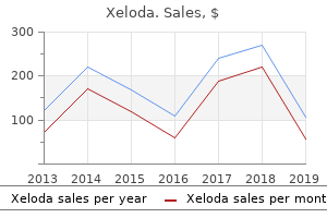
The box and whisker plots use a methodology which is unrelated to the method determining the map and column chart shading breast cancer awareness quotes xeloda 500mg line. They represent the data value at key rank positions when the geographical areas are rank-ordered according to data value size leading women's health issues purchase xeloda 500mg on-line. This graphic shows how variable the indicator is across all of the geographical areas womens health big book of exercises cheap 500mg xeloda amex. A single box and whisker plot is displayed for each time period so that comparisons can be made through time of the level and spread of values womens health 49 purchase xeloda 500 mg line. Overdispersion typically occurs when there are factors influencing the values that have not been accounted (or adjusted) for in the method of calculating the statistic, such as demographic risk factors, casemix or localised service configuration, which is particularly relevant to specialised services. These factors may account for the larger than expected number of areas with values greatly different from the England value. Wherever possible statistics presented in this Atlas have been adjusted for known influences, such as locality based variations in age structure, using techniques such as standardisation (see below). It is important to consider whether all known warranted factors have been adjusted for when assessing whether the observed variation is unwarranted. Box and whisker plots split the data presented into four equal parts in terms of the number of data points represented. Twenty-five per cent of data points lie between the maximum and the upper quartile, 25% of data points lie between the upper quartile and the median, 25% of data points lie between the median and the lower quartile, and 25% of data points lie between the lower quartile and the minimum. A box and whisker plot enables the user to obtain information about the shape or spread of the data points and in particular, whether or not the data points have a symmetric or skewed distribution. A dataset with a normal distribution is symmetric (non-skewed) around the mean (average), the mean and the median are equal to each other, and each half of the distribution is a mirror-image of the other half. In a distribution that is skewed there is a lack of symmetry between the upper and lower halves of the dataset. However, extreme outliers can heavily influence this 2010/11 2011/12 2012/13 2013/14 2014/15 statistic and consequently mislead about the extent of variability across the majority of the dataset. It may therefore be more helpful to use the 95th to 5th percentile (see below) 150 25 100 5 50 Min 5th percentile (the data value that lies in the 5% highest rank position) 0 Example 2005/06 Minimum (or smallest and therefore lowest ranked data point) 2006/07 2007/08 2008/09 2009/10 Inside the box is a horizontal line, which shows where the median (or Q2) lies. This statistic indicates the dispersion or spread of the data for the middle 50% of values. If there is an even number of values the median will be the average of the two central data points. Standardisation Differences in the number of events, for example incidence of disease, can be strongly related to the age structure of that population. In an attempt to identify variation that is beyond that related to different patterns of need, a technique called standardisation is used. This enables the level of testing to be compared between populations with different demographic structures producing a more level playing field. For instance if we compare two population groups, A and B, and population A has a higher rate of deaths when compared with population B we could conclude that population A has worse mortality outcomes in comparison with population B. However, if population A has a much higher proportion of older people in it we would expect population A to have a higher mortality rate when compared with population B because mortality rates are linked to increasing age. Therefore, it would be misleading to infer that people in population A are dying at a faster rate than people in population B. There are two main methods of calculating standardised rates: · · direct standardisation indirect standardisation · · the final column of the table is a summary of whether each of these four statistics is narrowing or widening (or median increasing/decreasing) and whether the trend is statistically significant at the 95% level. The statistical significance was determined using a two-tailed t-test on the slope of a linear regression line fitted to the values in the table over time, where the null hypothesis is that the slope equals zero. The significance test is only performed for indicators with data at three or more time periods. This regression line and the detailed results of the t-test are not presented in this Atlas. Only direct standardisation has been used within this Atlas and so only this method is discussed here. Directly standardised rates may adjust for the differences in age and sex distribution in a population and are usually expressed, for example, as a number of infections per 100,000 population. These calculations are then summed Data frequency the data frequency, ie the length of the time period for which data is presented, directly affects the number of observations represented in the visualisations. Statistical power, ie the ability to detect true differences, tends to increase with an increasing number of observations. This method of direct standardisation has been used for Maps 1a-c, 2, 4a-c, 19a, and 20.
Small bowel wall function in patients with advanced liver cirrhosis and portal hypertension: Studies on permeability and luminal bacterial overgrowth breast cancer stage 0 survival rate buy xeloda 500mg free shipping. Diagnosis of small intestinal overgrowth in patients with cirrhosis of the liver: Poor performance of the glucose breath hydrogen test menstrual uterine contractions cheap xeloda. Principles and clinical application of ultrasound elastography for diffuse liver disease Woo Kyoung Jeong1 women's health center queens ny generic xeloda 500mg amex, Hyo K menstruation pregnancy buy generic xeloda 500mg. Because of the inherent limitations of liver biopsy, there is a great need for noninvasive and reliable tests that accurately estimate the degree of liver fibrosis. Keywords: Elasticity imaging techniques; Liver cirrhosis; Hypertension, portal; Ultrasonography Received: January 9, 2014 Revised: March 9, 2014 Accepted: March 24, 2014 Correspondence to: Hyo K. The very small size of samples obtained through biopsy may not represent a heterogeneous distribution of liver fibrosis due to sampling bias [3]. Principles and clinical application of ultrasound elastography for diffuse liver disease. According to previous research on chronic hepatitis C, agreement among pathologists regarding the fibrosis grade is not excellent (about 0. Although the rate of complications is, very low and the risk has declined with the use of ultrasonographic guidance [6], liver biopsy is somewhat invasive and post-biopsy bleeding can be serious. With respect to non-invasive alternatives to liver biopsy, several serological or biochemical methods for the estimation of liver fibrosis have been validated primarily in patients with chronic hepatitis C, but still lack the ability to identify and classify the intermediate stages of fibrosis [7]. Introduced in 1991, elastography is another non-invasive technique for evaluating the elastic properties of soft tissue either quantitatively or qualitatively [8]. The elastography of the liver is theoretically not easy to determine compared with that of superficial organs because the liver is located deep and under the rib cage. The order of magnitude of the elastic modulus is approximately five times larger than that for other imaging modalities [9], meaning that the use of the elastic modulus can maximize the discrimination between different tissues or between normal tissue and lesions. The elastic modulus is defined as the slope of the stress-strain curve during elastic deformation. There are various approaches to elastic imaging, all of which consist of three basic steps: excitation (stress) application, tissue response (strain) measurement, and mechanical parameters estimation [9]. Excitation Application In its most basic form, shear wave-based elastography applies a perpendicular stress force on the target organ to induce "shear" on the tissue. By definition, shear is the change of shape (displacement)-without a change in volume-produced by a pair of forces acting in opposite directions. In the case of transient elastography, a mechanical push is used for excitation application, which produces transient shear waves in the target tissue. This type of excitation application is classified as a dynamic elastography technique with an external source. On the other hand, real-time tissue elastography methods are derived from the static elastography technique used for the measurement of breast tissue elasticity, and employ the quasi-static or intrinsic stress derived from heartbeats [12,13]. Through either a mechanical push or acoustic radiation force, the A-axis (direction of force=depth direction) displacement of the target tissue occurs, and the shear waves are generated simultaneously. Using the tissue displacement maps obtained during the period of shear wave propagation. In this way, the elastic modulus can be calculated by E=3 s2 where denotes the density of the tissue and Vs V represents the velocity of the shear wave. Tissue Response Measurement Measurement of tissue response is the most critical component of elastography. The basic measurement method consists of a Mechanical Parameter Estimation Both qualitative and quantitative methods are used to perform mechanical parameter estimation. Shear wave-based elastography applies a perpendicular stress force to a target organ in order to induce shear on the tissue. The information on the propagating shear wave including the velocity of the shear wave could be measured by obtaining radiofrequency images with a high frame rate, which can be used to generate a tissue displacement map. Then, the elastic property for quantitative estimation is calculated by the propagating velocity of the shear wave. The elastic properties for quantitative estimation are expressed as Young modulus (E) or the shear modulus. For most soft tissues, Young modulus and the shear modulus are related by a simple scale factor of 3: i. Relative tissue elasticity is thus calculated and displayed as a color overlay of the conventional B-mode image, and the strain ratio between two different points can be obtained instead of elastic modulus or shear wave velocity. Methods of Shear Wave-based Elastography Transient Elastography Transient elastography was the first commercialized elastography method developed to noninvasively assess the stiffness of deep soft tissues such as the liver.
With current therapy for the disease and the success of renal transplantation women's health equity act xeloda 500mg generic, however menopause 55 generic xeloda 500 mg without prescription, most patients live well into adulthood women's health clinic boca raton xeloda 500 mg with visa. Suspicion must be high in patients who present with diffuse symptoms menstrual 4 days late order 500 mg xeloda mastercard, particularly adolescent girls. It is characterized by activation of T and B lymphocytes, leading to vasculitis affecting small vessels of skeletal muscle, with immune complex deposition and subsequent inflammation of blood vessels and muscle. Initial use of pulse methylprednisolone and highdose oral prednisone (up to 2 mg/kg) frequently is required, followed by cautious tapering to minimize recurrence of symptoms. Hydroxychloroquine is used not only for the treatment of lupus skin disease, such as discoid lupus, but as maintenance therapy. Hydroxychloroquine treatment results in longer periods of wellness between flares of disease as well as decreased numbers of flares. Corticosteroids and hydroxychloroquine frequently are not sufficient therapies for lupus nephritis or cerebritis. Cyclophosphamide is effective for the worst forms of lupus nephritis, with significant improvements in outcome and decreased rates of progression to renal failure. For patients who are not able to tolerate the tapering of corticosteroids, the use of steroid-sparing agents, such as azathioprine, methotrexate, or mycophenolate mofetil, may be indicated. Because of this prohibition, patients benefit from calcium and vitamin D supplementation to reduce the risk of osteoporosis that may result from prolonged corticosteroid use. Early treatment of hyperlipidemia to decrease long-term cardiovascular complications is also indicated. Dermatomyositis tends to present in a slow, progressive fashion, with insidious onset of fatigue, malaise, and progressive muscle weakness, accompanied by low-grade fevers and rash. Some children present in an acute fashion, however, with rapid onset of severe disease. Children have difficulty climbing steps, getting out of chairs, and getting off the floor. In severe cases, the patient is not able to sit up from a supine position or even lift the head off the examination table (see Chapter 182). Scaly, red plaques (Gottron papules) classically are found across the knuckles but can be found on the extensor surfaces of any joint. Less commonly, patients develop cutaneous vasculitis, with inflammation, erythema, and skin breakdown. Dystrophic calcification can occur in the skin and soft tissues in any area of the body; it ranges from mild to extensive (calcinosis universalis). Although it is difficult to predict who will develop calcinosis, it occurs more commonly in children with cutaneous vasculitis, prolonged disease activity, or delays in onset of therapy. Control of insulin resistance frequently leads to improvement in muscle disease in these patients. Patients who ultimately develop calcinosis are at risk for chronic loss of mobility, depending on the extent of calcium deposition. The association of dermatomyositis with malignancy seen in adults does not occur in children. A small percentage of children have muscle disease without skin manifestations, but polymyositis is sufficiently rare in children that they should have a muscle biopsy to exclude other causes of muscle weakness, such as muscular dystrophy (particularly boys). The differential diagnosis also includes postinfectious myositis and other myopathies (see Chapter 182). The peak age range is 3 to 7 years; the syndrome seems to be more common in boys than girls. Children with growing pains complain of deep, crampy pain in the calves and thighs. It most typically occurs in the evening or as nocturnal pain that occasionally can waken the child from sleep. Growing pains tend to be more common in children who are extremely active; bouts are exacerbated by increased physical activity. The physical examination is unremarkable, with no evidence of arthritis or muscular tenderness or weakness.
Buy xeloda pills in toronto. National Meeting on Active Duty and Vet Womens Health Part 1 Overview.
St. Augustine Humane Society | 1665 Old Moultrie Rd. | St. Augustine, FL 32084 PO Box 133, St. Augustine, FL 32085 | Phone (904) 829-2737 |info@staughumane.org
Hours of Operation: Mon. - Fri. 9:00am - 4:00pm Closed for Lunch Each Day: 12:30pm - 1:30pm
Open Sat. by Appointment Only for Grooming General Operations Closed: Sat. and Sun.